No products
Prices are tax excluded
Product successfully added to your shopping cart
There are 0 items in your cart. There is 1 item in your cart.
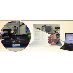
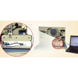
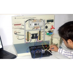
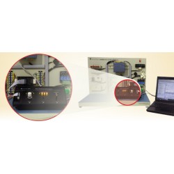
Scientech11E Ultrasonic Investigation with the Eye Dummy
Scientech11E
New
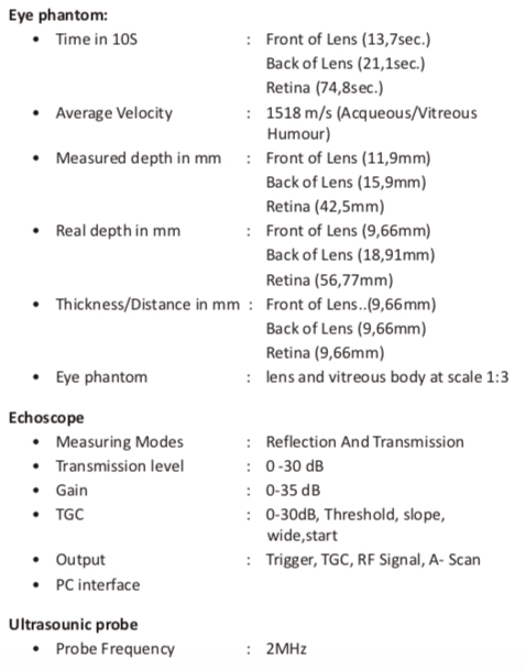
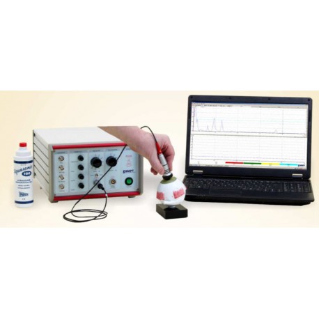
In this experiment a typical application of A-Scan ultrasound biomatry diagnostics used in ophthalmology is given. At an eye dummy all parts of the healthy eye are mearused and correction shell be done.
- Consulta este producto
- Remove this product from my favorite's list.
- Add this product to my list of favorites.
| Category | Biomedicine |
Scientech11E Ultrasonic Investigation with the Eye Dummy is an ideal platform to enhance education, training, skills & development amongs our young minds.
- Ultrasonic investigation with the eye dummy
Ultrasound is used also in ophthlmology. Its largest importance lies in the area of biometry, in the measurement of distance in eye. The distance between cornea and retina is very significant for the calculation of the characteristics of the artificial lens implanted to patients with cataract. Sonography is necessary in this case since the cornea or the lens are too cloudy for the used of optical methods. Investigations of the aqueous, vitreous humour and the thickness of the lens are nowadays often done with new methods of laser light or ultrasonic B-mode imaging. The given measurement time of flight of the echoes, the A-scan can not be calculate as distance in a simple way, because of different velocity in the different media (cornea, lens, vitreous humour). Therefore a corrective calculation is necessary.Two velocity are given for the dummy:
-Lens: 2500 m/s
-Humour: 1410 m/s
These values and the time of flight from the measured A-scan image shall be used to determine the distances with the help of the following equations
(1) S = ν (Δt/2)
In medical diagnostics averages are often used known from experience. This average velocity shall be calculated for the dummy with the following equation.
(2) ν = ν1 (t1+(t3 - t2) + v2 (t2 - t1) / t3
- Procedure
Ultrasonic coupling gel is used to connect the probe to the cornea of the dummy. Slowly move the probe over the cornea to look for the optimal signal (2 large lens peaks and one smaller from the retina). After measuring the time of flight of the peaks the real distances can be calculated. ​
